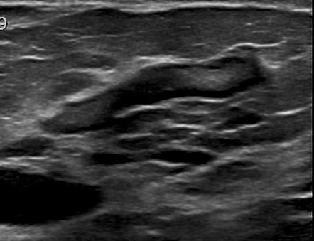
Anatomy Of Chest
The chest or thorax is the region between the neck and diaphragm that encloses organs, such as the heart, lungs, esophagus, trachea, and thoracic diaphragm. Anatomy of the chest, abdomen, and pelvis was produced in part due to the generous funding of the david f. Plus, how to target each to make them bigger and stronger. 20/01/2018 · the dominant muscle in the upper chest is the pectoralis major. 15/10/2017 · radiology basics of chest ct anatomy with annotated coronal images and scrollable axial images to help medical students and junior doctors learning anatomy.

Swensen fund for innovation in teaching.
The chest or thorax is the region between the neck and diaphragm that encloses organs, such as the heart, lungs, esophagus, trachea, and thoracic diaphragm. Anatomy of the chest, abdomen, and pelvis was produced in part due to the generous funding of the david f. 20/01/2018 · the dominant muscle in the upper chest is the pectoralis major. 15/10/2017 · radiology basics of chest ct anatomy with annotated coronal images and scrollable axial images to help medical students and junior doctors learning anatomy. Anatomy of the thorax, heart, abdomen and pelvis recommended text gray's anatomy for students. 14/09/2015 · the chest anatomy includes the pectoralis major, pectoralis minor and the serratus anterior. Find out more about the individual muscles within the chest anatomy by clicking their respective. This mri chest (thorax) axial cross sectional anatomy tool is absolutely free to use. Of the two chest muscles, the pectoralis major (a.k.a. The pec major) is the one that commands the most real estate. Other important structures, such as the pleura, only become visible when abnormal, and some are not visible at all, such as the phrenic nerve. 16/07/2019 · in addition to moving the arm and pectoral girdle, muscles of the chest and upper back work together as a group to support the vital process of breathing. Computed tomography (ct) of the chest can detect pathology that may not show up on a conventional chest radiograph (1).
This page provides an overview of the chest muscle group. The chest or thorax is the region between the neck and diaphragm that encloses organs, such as the heart, lungs, esophagus, trachea, and thoracic diaphragm. Of the two chest muscles, the pectoralis major (a.k.a. Find out more about the individual muscles within the chest anatomy by clicking their respective. Computed tomography (ct) of the chest can detect pathology that may not show up on a conventional chest radiograph (1).

14/09/2015 · the chest anatomy includes the pectoralis major, pectoralis minor and the serratus anterior.
Computed tomography (ct) of the chest can detect pathology that may not show up on a conventional chest radiograph (1). 16/07/2019 · in addition to moving the arm and pectoral girdle, muscles of the chest and upper back work together as a group to support the vital process of breathing. 20/01/2018 · the dominant muscle in the upper chest is the pectoralis major. Find out more about the individual muscles within the chest anatomy by clicking their respective. Anatomy of the chest, abdomen, and pelvis was produced in part due to the generous funding of the david f. Swensen fund for innovation in teaching. The pec major) is the one that commands the most real estate. 15/10/2017 · radiology basics of chest ct anatomy with annotated coronal images and scrollable axial images to help medical students and junior doctors learning anatomy. This page provides an overview of the chest muscle group. This mri chest (thorax) axial cross sectional anatomy tool is absolutely free to use. Of the two chest muscles, the pectoralis major (a.k.a. Other important structures, such as the pleura, only become visible when abnormal, and some are not visible at all, such as the phrenic nerve. Use the mouse scroll wheel to move the images up and down alternatively use the tiny arrows (>>) on both side of the image to move the images.>>) on both side of the image to move the images.
20/01/2018 · the dominant muscle in the upper chest is the pectoralis major. 29/08/2020 · here, we break down the anatomy of your chest muscles. Swensen fund for innovation in teaching. Anatomy of the thorax, heart, abdomen and pelvis recommended text gray's anatomy for students. Computed tomography (ct) of the chest can detect pathology that may not show up on a conventional chest radiograph (1).

Plus, how to target each to make them bigger and stronger.
This page provides an overview of the chest muscle group. The chest or thorax is the region between the neck and diaphragm that encloses organs, such as the heart, lungs, esophagus, trachea, and thoracic diaphragm. 14/09/2015 · the chest anatomy includes the pectoralis major, pectoralis minor and the serratus anterior. Anatomy of the thorax, heart, abdomen and pelvis recommended text gray's anatomy for students. Learn about each of these muscles, their locations, functional anatomy and exercises for them. The sternum is also included on a frontal view but it overlies other midline structures and so is obscured. Use the mouse scroll wheel to move the images up and down alternatively use the tiny arrows (>>) on both side of the image to move the images.>>) on both side of the image to move the images. Other important structures, such as the pleura, only become visible when abnormal, and some are not visible at all, such as the phrenic nerve. Find out more about the individual muscles within the chest anatomy by clicking their respective. Of the two chest muscles, the pectoralis major (a.k.a. Plus, how to target each to make them bigger and stronger. 15/10/2017 · radiology basics of chest ct anatomy with annotated coronal images and scrollable axial images to help medical students and junior doctors learning anatomy. Computed tomography (ct) of the chest can detect pathology that may not show up on a conventional chest radiograph (1).
Anatomy Of Chest. 16/07/2019 · in addition to moving the arm and pectoral girdle, muscles of the chest and upper back work together as a group to support the vital process of breathing. 29/08/2020 · here, we break down the anatomy of your chest muscles. Computed tomography (ct) of the chest can detect pathology that may not show up on a conventional chest radiograph (1). Of the two chest muscles, the pectoralis major (a.k.a. Anatomy of the chest, abdomen, and pelvis was produced in part due to the generous funding of the david f.
Posting Komentar untuk "Anatomy Of Chest"